WHAT IS MAGNETIC RESONANCE IMAGING OF THE HEAD OR BRAIN AND HOW DOES IT WORK?

If you've ever heard of head or brain MRI and wondered how this type of study works, you're in the right place.
This guide provides a clear and comprehensive explanation of what a head MRI is, how it functions, and why it has become a cornerstone in medical diagnostics.
By the end of this article, you'll understand how this procedure helps diagnose a wide range of conditions and the key differences between an MRI and other imaging studies like CT scans.
How does MRI of the head or brain work?
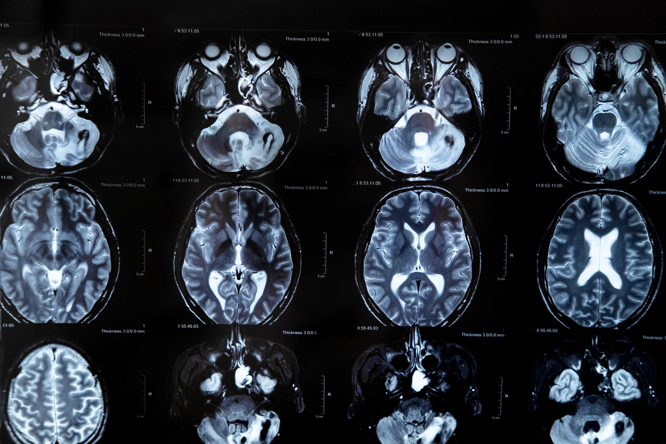
A head MRI, also referred to as a brain MRI, is an imaging test that provides highly detailed views of brain tissues and structures.
It uses powerful magnetic fields and radio waves to create detailed images without the need for radiation. This makes the procedure particularly safe and effective in detecting abnormalities such as small tumors, bleeding, or complex neurological disorders.
During a head MRI, the patient lies inside an MRI machine, which generates a magnetic field that temporarily aligns the protons in the body’s cells.
Radio waves then stimulate these protons, producing signals that are captured and converted into detailed images of the brain.
Differences between head or brain MRI and cranial CT scanning
It is common for people to confuse head or brain MRI with cranial computed tomography (CT). It is important to emphasize that these are two imaging techniques used to examine the brain and skull, but they have fundamental differences in terms of their principle of operation, advantages, limitations and applications.

What to expect in the process of performing an MRI of the head or brain?
- The procedure lasts between 30 to 60 minutes.
- You’ll lie on a table with your head positioned inside the machine.
- It’s crucial to remain still during the scan.
- The machine will produce loud noises, but you’ll be provided with earplugs or headphones.
- If contrast is used, a liquid will be injected through a vein in the arm.
- The procedure is painless and safe.
Myths and truths about MRI of the head or brain
With many misconceptions about MRIs, it’s easy to have doubts or concerns. Let’s clarify some of the most common myths:
- Myth 1: A head MRI damages the brain.
Fact: MRIs are entirely safe. They do not use radiation and have no harmful effects on the brain or body. - Myth 2: MRIs are painful or invasive.
Fact: MRIs are non-invasive and painless. While some patients may feel discomfort due to the noise or enclosed space, many facilities offer headphones or other solutions for comfort. - Myth 3: I can’t have an MRI if I have metal implants.
Fact: Not all implants prevent an MRI, but some may interfere. It’s essential to inform the medical team about any metal in your body to ensure safety.
Have more questions about this procedure? Check out related articles like Brain MRI: Understanding the Procedure to learn how these advancements improve diagnostic accuracy.
Conclusion: Importance of head or brain resonance in medical diagnosis.
MRI of the head or brain is an invaluable tool in the diagnosis of neurological problems. With its ability to show precise details of the brain and other internal structures, it is essential for detecting, monitoring and treating health conditions early and effectively.
If you have neurological symptoms, a family history of brain conditions, or simply want to learn more about this procedure, consult your doctor to determine if a head MRI is right for you.
Visit our website for detailed information and find out how we can help you take care of your well-being.