What Is a Contrast-Enhanced Brain MRI, and When Is It Necessary?
Contrast Brain MRI is a key procedure in the diagnosis of various brain conditions. In this blog, we explain what this study consists of, when it is necessary and the benefits it offers to obtain more accurate and complete diagnoses. If you are looking for information about this procedure, you have come to the right place.
By using a contrast agent, this test provides more detailed images that can reveal abnormalities not visible on a traditional MRI scan.
What is a contrasted brain MRI?
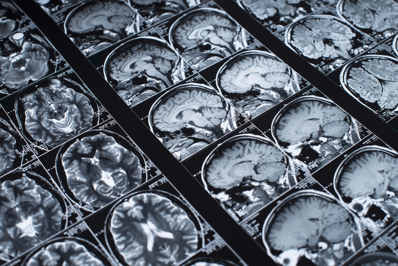
A contrast-enhanced brain MRI is an imaging test that uses a special contrast agent, typically gadolinium-based, to highlight specific details in the brain.
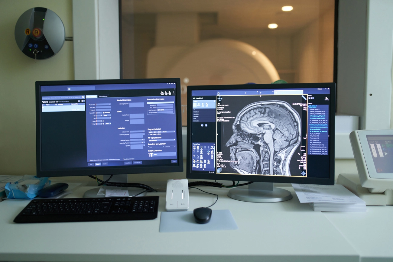
This procedure is performed in an MRI machine and allows radiologists to view areas of the brain with enhanced clarity, identifying issues that might otherwise be overlooked.
When the contrast agent is injected into the patient’s bloodstream, it travels to the brain, highlighting blood vessels, tumors, and areas of inflammation or damage. This process is vital for detecting conditions like tumors, brain injuries, and other complex disorders.
When is a contrasted brain MRI needed?
A contrast-enhanced brain MRI is recommended when a deeper analysis is required for certain conditions. Some of the critical scenarios include:
- Detecting tumors or abnormal masses: The contrast helps differentiate healthy tissues from suspicious growths.
- Studying neurological diseases: Conditions such as multiple sclerosis, aneurysms, and specific infections demand detailed imaging only achievable with contrast.
- Evaluating inflammation or infections: For cases like encephalitis or brain abscesses, contrast highlights inflamed areas indicative of infections.
- Neurological disorders: To detect causes of persistent headaches, memory loss or cognitive problems.
This test is also suggested when abnormalities are detected in a non-contrast MRI, as the contrast provides a more comprehensive and precise view of brain tissues.
What is the difference between contrasted and non-contrasted MRI?
The primary distinction between contrast-enhanced and non-contrast brain MRIs lies in the clarity and visibility of the results. While a non-contrast MRI provides a general overview of the brain, a contrast-enhanced MRI delivers a more detailed view of the tissues.
- In non-contrast MRI, only brain structures are visualized without highlighting certain areas, which may be suitable for evaluating more general conditions.
- Contrast-enhanced MRI makes it possible to highlight specific areas of the brain, such as tumors or blood vessels, which significantly improves diagnostic accuracy. Contrast makes the images much more detailed, allowing the detection of smaller abnormalities that might otherwise go undetected in a non-contrast study.
Characteristics of non-contrast magnetic resonance imaging:
- Non-invasive, as no additional substances are required.
- Ideal for initial and routine imaging.
- May be less effective in identifying certain masses or tumors.
Characteristics of magnetic resonance imaging with contrast:
- Uses gadolinium, a contrast agent injected into the bloodstream to improve image clarity.
- Essential for detailed diagnoses and monitoring of complex conditions.
- Provides precise information on blood flow, inflammation, and tumors.
The choice between the two depends on the required diagnosis, patient symptoms, and the physician’s recommendations.
Benefits and precautions of contrasted MRI brain imaging
Benefits:
- Early and accurate diagnosis: Contrast allows detection of abnormalities and lesions at early stages, crucial for severe conditions.
- Enhanced detail: The added clarity from contrast helps identify small tumors, infections, and other issues.
- Diagnostic versatility: It is a key tool for diagnosing complex conditions like multiple sclerosis and vascular diseases.
Precautions:
- Allergic reactions: Although rare, some individuals may experience allergic reactions to gadolinium.
- Kidney function: Patients with kidney issues should be evaluated beforehand, as gadolinium may pose risks.
- Metal in the body: Patients with metallic devices or implants in the body, such as pacemakers, should consult with their physician before undergoing an MRI.
- Claustrophobia: Some individuals may feel discomfort during the procedure, which requires staying in the MRI machine for an extended period.
At HCI, we ensure the procedure is safe and effective by carefully assessing patients and offering support throughout the process. We have a highly trained team ready to assist and support you every step of the way. From preparation to interpretation of the results, we are here to make you feel at ease and in good hands. Your health is our priority and we are committed to providing you with the best care at all times!
Conclusion: The importance of a contrasted brain MRI in medical diagnosis.
A contrast-enhanced brain MRI is an invaluable tool in modern medicine. Its ability to provide detailed images and identify complex conditions makes it essential for accurate and timely diagnoses.
If you’re experiencing neurological symptoms, have a family history of brain conditions, or need further information, consider consulting with a specialist to determine if a contrast-enhanced brain MRI is right for you.
If you need more information or would like to make an appointment, don't hesitate to contact us, we are here to help you!
Want to learn more? Visit our website to discover how this diagnostic tool can help you take better care of your health.